What Is Fragmentomics?
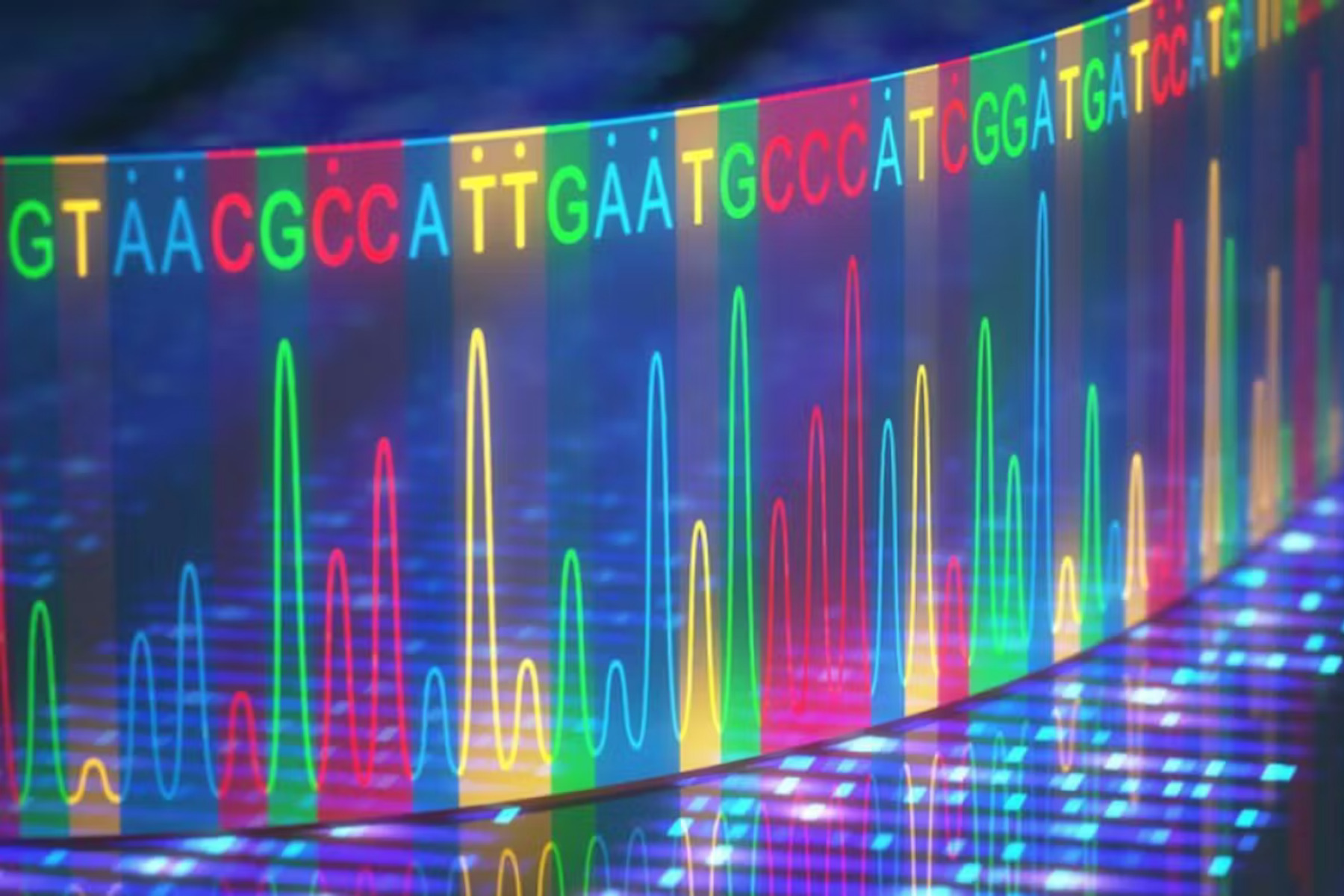
In recent years, researchers have realized that circulating cell-free DNA (cfDNA) in the blood carries far more information than just mutations. cfDNA fragments reflect the way chromatin is packaged and cut during apoptosis. Their sizes, genomic locations, and end motifs form recognizable “fragmentation patterns,” collectively called fragmentomics. These patterns preserve the nucleosomal structure of the tissues that released them, providing a type of epigenetic fingerprint. Importantly, cancers show distinctive fragmentation because chromatin and transcriptional regulation are altered in tumors [1,2].
The ability to noninvasively interrogate cfDNA fragmentation means that fragmentomics could help detect cancer earlier, track its course, and complement existing genomic methods. The approach belongs to the family of “liquid biopsies,” which aim to provide molecular insights from a simple blood draw.
MRD and Its Clinical Importance
Minimal residual disease (MRD) refers to the tiny number of cancer cells or their molecular traces that persist after treatment. While invisible on imaging, they can later drive relapse. MRD monitoring has become standard in many hematological cancers, and interest in applying it to solid tumors has accelerated.
Evidence shows that detecting ctDNA after surgery can reliably predict recurrence. In stage II colon cancer, patients with ctDNA positivity had relapse rates exceeding 80% [3], and similar findings were reported in early-stage lung cancer [4]. A randomized trial even demonstrated that ctDNA-informed decisions could safely reduce chemotherapy exposure in stage II colon cancer without worsening survival [5].
The clinical stakes are high: patients who are truly free of disease could be spared toxic treatments, while those with detectable MRD might benefit from intensified or prolonged therapy.
How Fragmentomics Enhances MRD Detection
Most current MRD assays track tumor-specific mutations identified from sequencing the patient’s tumor tissue. These tests are highly specific but can lack sensitivity, especially when ctDNA levels are extremely low immediately after surgery or when the tumor harbors few mutations. They also require prior access to tumor tissue, which is not always available.
Fragmentomics provides a tumor-naïve, plasma-only strategy. Instead of searching for predefined mutations, it examines global fragmentation features across the genome. Machine learning models can then classify whether a cfDNA profile is consistent with residual cancer [6,7].
This approach has shown substantial promise. In non-small cell lung cancer, integrating fragmentomics with mutation detection improved sensitivity for identifying patients at risk of recurrence, raising detection from ~43% with mutations alone to ~78% when both signals were combined [8]. Importantly, the method remained effective even in “low-shedding” cancers where mutation-based ctDNA was almost undetectable.
Moreover, fragmentomics can be layered with other epigenetic readouts, such as DNA methylation, to further strengthen detection accuracy. Studies suggest that multi-omic assays could ultimately achieve both high sensitivity and high specificity [9].
How Fragmentomics Enhances MRD Detection
- Earlier and more sensitive alerts. By capturing subtle genome-wide patterns, fragmentomics increases the chances of identifying MRD when ctDNA concentrations are below the detection threshold of mutation-only assays [8].
- Tumor-agnostic scalability. Because fragmentomics does not require tumor sequencing, it simplifies logistics and accelerates testing. Plasma-only workflows are particularly useful for community settings where access to tissue samples is limited [6,7].
- Versatility. Fragmentomics has applications beyond MRD, including early cancer screening, prenatal testing, monitoring transplant rejection, and even infectious disease surveillance [3].
- Pathway to next-generation MRD assays. The integration of fragmentomics with mutation and methylation analysis is likely to define the next era of liquid biopsy, supporting personalized cancer care and adaptive treatment strategies[9].
Conclusion
Fragmentomics represents a significant leap in how we interpret the information hidden within cfDNA. By focusing on how DNA fragments are cut and distributed, rather than only on mutations, fragmentomics provides a complementary and often more sensitive signal for minimal residual disease detection. In cancers such as lung and colorectal, where relapse risk remains high even after curative-intent surgery, fragmentomics can deliver earlier warnings and more accurate prognoses.
The field is still young, but the direction is clear: combining fragmentation patterns with other molecular features will enable highly sensitive, tumor-agnostic, and clinically practical MRD tests. This convergence of biology, sequencing, and machine learning could transform how clinicians monitor patients, personalize therapies, and ultimately prevent cancer from returning.
Literature
- Snyder MW, Kircher M, Hill AJ, et al. Cell-free DNA Comprises an In Vivo Nucleosome Footprint that Informs Its Tissues-Of-Origin. Cell. 2016;164(1–2):57–68.
- Cristiano S, Leal A, Phallen J, et al. Genome-wide cell-free DNA fragmentation in patients with cancer. Nature. 2019;570(7761):385–389.
- Tie J, Wang Y, Tomasetti C, et al. Circulating tumor DNA analysis detects minimal residual disease and predicts recurrence in patients with stage II colon cancer. Sci Transl Med. 2016;8(346):346ra92.
- Abbosh C, Birkbak NJ, Wilson GA, et al. Phylogenetic ctDNA analysis depicts early-stage lung cancer evolution. Nature. 2017;545(7655):446–451.
- Tie J, Wang Y, Lo SN, et al. Circulating tumor DNA analysis guiding adjuvant therapy in stage II colon cancer: 5-year outcomes of the randomized DYNAMIC trial. Nat Med. 2025;31(5):1509–1518.
- Parikh AR, Van Seventer EE, Siravegna G, et al. Minimal Residual Disease Detection using a Plasma-only Circulating Tumor DNA Assay in Patients with Colorectal Cancer. Clin Cancer Res Off J Am Assoc Cancer Res. 2021;27(20):5586–5594.
- Chan HT, Nagayama S, Otaki M, et al. Tumor-informed or tumor-agnostic circulating tumor DNA as a biomarker for risk of recurrence in resected colorectal cancer patients. Front Oncol. 2022;12:1055968.
- Wang S, Xia Z, You J, et al. Enhanced Detection of Landmark Minimal Residual Disease in Lung Cancer Using Cell-free DNA Fragmentomics. Cancer Res Commun. 2023;3(5):933–942.
- Nguyen VTC, Nguyen TH, Doan NNT, et al. Multimodal analysis of methylomics and fragmentomics in plasma cell-free DNA for multi-cancer early detection and localization. Choi M, editor. eLife. 2023;12:RP89083.


